
In Search of Solutions
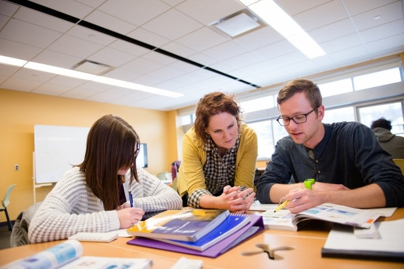
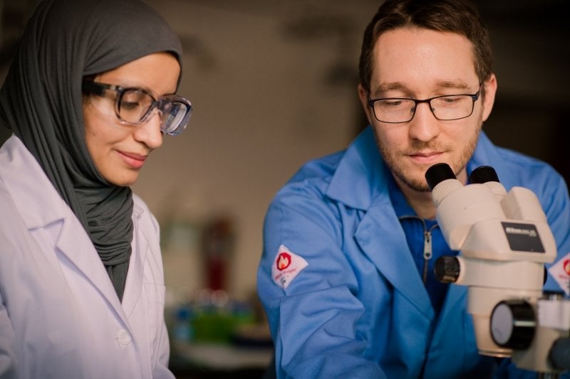
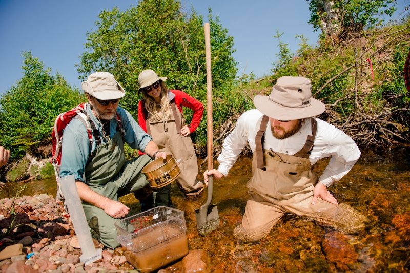
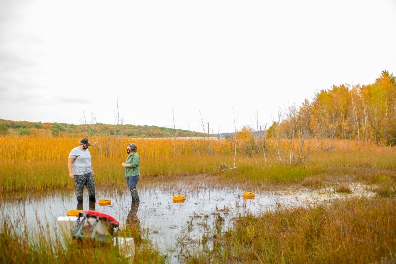
Biological Sciences brings together students and faculty in search of solutions to biological problems—from medicine to freshwater ecosystem health. Use molecular tools to grow, modify, and characterize plants, animals, and microorganisms in the lab. Present results in classrooms, research symposia, and scientific journals. Michigan Tech students work and play in all seasons of our outdoor playground, the Keweenaw Peninsula.
Diversity, Equity and Inclusion
We believe in the strength of diversity!
The biological sciences link all aspects of life, reaching not only across scales from molecules, to cells, to ecosystems, but also across cultural and social identities. Therefore, excellence in the biological sciences requires the fostering of diversity and equity across the discipline. The Department of Biological Sciences at Michigan Technological University is committed to creating a diverse, equitable, inclusive, and safe environment for all our faculty, staff, and students. This community approach will provide ample opportunities for everyone to realize their personal goals while contributing to the success of the department. We understand that much work needs to be done, and we all pledge to work together to build and support a culture and community where everyone is respected and welcomed.
Open Positions
For Faculty
For Students
- Dr. Yan Zhang's Lab - Ph.D. Research Assistantship
- Dr. Trista Vick-Majors' Lab - Microbial Ecology and Winter Limnology
- Dr. Gordon Paterson's Lab - Aquatic Ecotoxicology and Ecology
- Dr. Erika Hersch-Green's Lab - Postdoctoral Researcher Opportunity in Plant Evolutionary Ecology
- Dr. Erika Hersch-Green's Lab - REU Internship in Plant Ecology and Evolutionary Biology
- Dr. Erika Hersch-Greens's Lab - PhD Student Opportunity to Join Dr. Erika Hersch-Green’s Research Group in Plant Evolutionary Ecology
Ready for Medical School"I liked that my undergrad degree was so diverse. In addition to biology classes, I also took lots of courses in chemistry, physics, writing, and even computer science, and it was really fun getting to explore all these different disciplines and challenge myself in new ways all the time. My major definitely kept me on my toes, which I know will help me during medical school." Read Elise's Advice to Pre-Med Students.
